Knee Joint, Osteoarthritis, 3D Joint Model
膝关节,骨关节,3D关节模型
English Prompt:
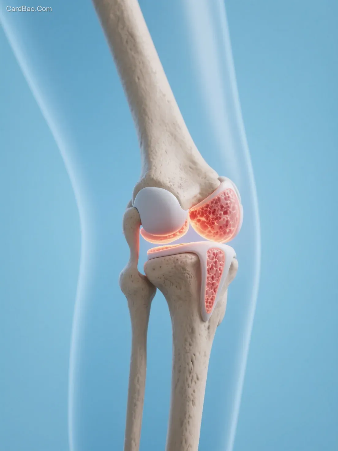
Knee joint, bone joint, 3D joint model. In terms of style, it adopts a medical visualization approach, utilizing precise and clear visual presentation to intuitively display the complex structure of the knee joint, combining scientific accuracy with educational value. Commonly used in medical education and health science communication scenarios.

Key elements include the anatomical structure of the knee joint, featuring bones such as the femur, tibia, and patella, as well as joint surfaces and possible bone details (e.g., micro-cracks simulating pathology). The overall visual language is realistic yet simplified, effectively conveying medical knowledge about the knee joint. Inflammatory areas such as the synovium and cartilage are highlighted.

The background is light blue, with a large scene composition, photography-style rendering, depth of field, high-definition quality, rich details, and a blurred background to simulate a real photographic setting.
中文Prompt:
膝关节,骨关节,3D关节模型,从风格看,是医学可视化风格,借助精准、清晰的视觉呈现,把复杂膝关节结构直观展现,兼具科学性与科普性,常用于医学教育、健康科普场景,元素方面,核心是膝关节解剖结构,呈现股骨、胫骨、髌骨等骨骼,还有关节面、可能的骨质细节(如细微裂纹模拟病理);整体通过写实又简化的视觉语言,传递膝关节医学知识,高亮显示炎症区域如滑膜、软骨,浅蓝色背景,大场景图,场景图,摄影,景深,高清,细节丰富,背景虚化,真实摄影场景。
Resolution: 1104×1472